Is It a Bad Fold or an Intrinsically Disordered Region? A Coarse-Grained MD Look at IFITM2
Posted: 2026-08-18
I recently found a computationally folded (with AlphaFold) transmembrane (TM) protein with a very poor fold (<70 pLDDT), interferon-induced transmembrane protein 2 (IFITM2), which is known to prevent the entry of Hepatitis C virus into the cell (Narayana et al., JBC. 2015). I wanted to see if I could refine the protein in the context of a lipid bilayer (simulating the cellular membrane) through a coarse-grained (CG) molecular dynamics (MD) simulation. IFITM2 is a 132 amino acid-long human antiviral protein, containing two ~10 amino-acid long predicted alpha-helices, one being a predicted transmembrane helix, and an N-terminus which appeared to be either a poorly folded region...or could it be an intrinsically disordered region or IDR? An IDR is a protein region that lacks structure, but has a function nonetheless (see Kjaergaard and Kragelund, Cell Mol Life Sci. 2017, for a good review and Dunker et al. IDP. 2013 for nomenclature).
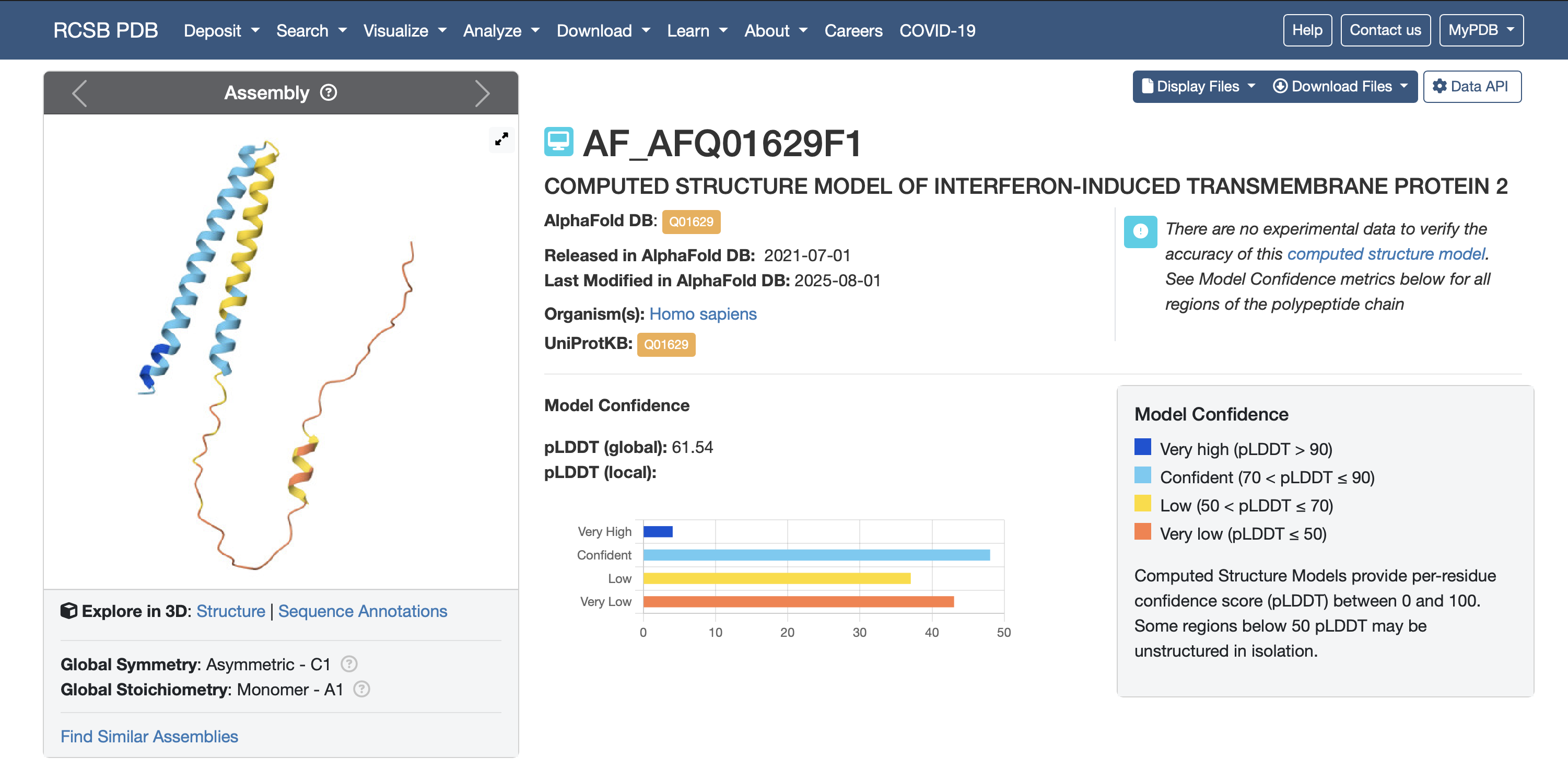
Wait…so this might not be a poor fold after all and this region is an intrinsically disordered region in the cell (even if disordered transiently)? I wanted to find out more if I could…I started reading papers on intrinsic disorder in membrane proteins and decided to perform some simple CG production/stability MD simulations (with proper energy minimization and NVT then NPT equilibration).
Two out of three 1 microsecond production CG MD simulations, with a pure POPC lipid bilayer, showed two C-terminal transmembrane helices and the N-terminal region associating with the membrane (on it, not in it), in what appeared to be an amphipathic manner (hydrophilic part on one side and hydrophobic part on the opposite side).
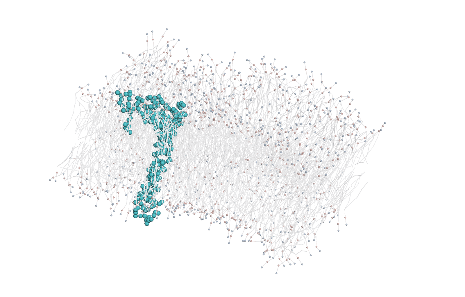
There was even a small helix in the secondary structure of that N-terminal region (predicted to be cytosolic - see UniProt entry Q01629), part of a possible amphipathic helix, one mechanism for disordered protein attachment to membranes (MacAinsh et al. Annu Rev Biophys. 2025). Interestingly, in the closely related IFITM3, a stretch long annotated as a transmembrane domain turned out to be a conserved amphipathic helix that is required for antiviral activity and that also induces negative membrane curvature (Chesarino et al. EMBO Rep. 2017), so I am now less sure as to if I can call both of the C-terminal helices transmembrane. Could these also be part of a signaling pathway?
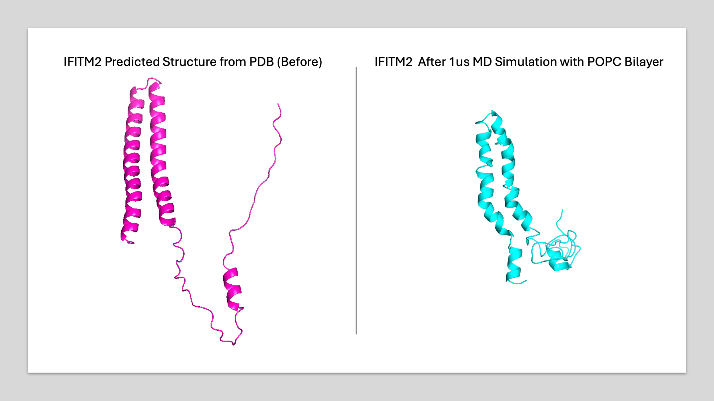
What could this mean for my questions - did I refine the structure correctly (my potential energy appeared plausible throughout the simulation)? Is this actually an IDR with some dynamic and real behavior in the cell; or is this an amphipathic domain/helix that helps anchor the protein with possibly some other cellular function? It is known that IDRs have a prevalence of secondary-structure-destabilizing amino acid residues like proline and glycine as well as repeat motifs and prevalence of PTMs (Kjaergaard and Kragelund, Cell Mol Life Sci. 2017). Indeed this N-terminus has an abundance of prolines as compared to the rest of the sequence (15.6% of residues 1-45 versus 2.3% across the rest of the protein) as well as a possible phosphorylation site. And that site is not just any tyrosine: residues 19-22 are YEML, the AP-2 sorting motif that sends IFITM2 and IFITM3 into endosomes (Jia et al. Cell Microbiol. 2014), and phosphorylation of that tyrosine by Src kinases blocks both its endocytosis and its degradation (Chesarino et al. JBC. 2014).
The movie above was made from the MD simulation production structures (with the first frame being the predicted structure from the PDB), Python and an open-source version of PyMOL. Simulations were run in OpenMM (Eastman et al. The Journal of Physical Chemistry B. 2024) with the Martini 3 coarse-grained force field (Souza et al. Nature Methods. 2021), in a pure POPC bilayer.
So, is it a bad fold or an IDR?
I do not think 1 microsecond of coarse-grained sampling is enough to draw concrete conclusions, but the N-terminus did not behave like a modeling artifact. In two out of three replicates it strayed from the extended conformation AlphaFold had given it and settled compact and partly helical onto the cytosolic face of the bilayer, which looks a lot more like a disordered region folding upon binding to a membrane than like a simply bad prediction. Also, a low pLDDT is a confidence score and not a disorder prediction, even if the two may travel together.
This evidence is admittedly limited. Three replicates is a small number and one of them disagreed. A pure POPC bilayer is not a plasma membrane (no cholesterol, no anionic lipids), and I did not model the S-palmitoylation that the IFITM proteins need for their antiviral activity (Narayana et al. JBC. 2015).
So what next? I would like to run an actual disorder predictor over this sequence and see how it lines up with pLDDT, since at the moment my case for disorder leans on pLDDT plus amino acid composition. I would also like to see whether that small N-terminal helix survives at atomistic resolution, and to rerun all of this in a more realistic membrane with cholesterol and anionic lipids. Certainly many directions to go!
References
- Kjaergaard, M. & Kragelund, B. B. (2017) "Functions of intrinsic disorder in transmembrane proteins." Cellular and Molecular Life Sciences 74(17), 3205-3224. doi:10.1007/s00018-017-2562-5
- Dunker, A. K., Babu, M. M., Barbar, E., et al. (2013) "What's in a name? Why these proteins are intrinsically disordered." Intrinsically Disordered Proteins 1(1), e24157. doi:10.4161/idp.24157
- MacAinsh, M., Kunnath Muhammedkutty, F. N., Prasad, R. & Zhou, H.-X. (2025) "Membrane Association of Intrinsically Disordered Proteins." Annual Review of Biophysics 54(1), 275-302. doi:10.1146/annurev-biophys-070124-092816
- Chesarino, N. M., Compton, A. A., McMichael, T. M., et al. (2017) "IFITM3 requires an amphipathic helix for antiviral activity." EMBO Reports 18(10), 1740-1751. doi:10.15252/embr.201744100
- Narayana, S. K., Helbig, K. J., McCartney, E. M., et al. (2015) "The Interferon-induced Transmembrane Proteins, IFITM1, IFITM2, and IFITM3 Inhibit Hepatitis C Virus Entry." Journal of Biological Chemistry 290(43), 25946-25959. doi:10.1074/jbc.M115.657346
- Jia, R., Xu, F., Qian, J., et al. (2014) "Identification of an endocytic signal essential for the antiviral action of IFITM3." Cellular Microbiology 16(7), 1080-1093. doi:10.1111/cmi.12262
- Chesarino, N. M., McMichael, T. M., Hach, J. C. & Yount, J. S. (2014) "Phosphorylation of the Antiviral Protein Interferon-inducible Transmembrane Protein 3 (IFITM3) Dually Regulates Its Endocytosis and Ubiquitination." Journal of Biological Chemistry 289(17), 11986-11992. doi:10.1074/jbc.M114.557694
Structure prediction and visualization
- Jumper, J., Evans, R., Pritzel, A., et al. (2021) "Highly accurate protein structure prediction with AlphaFold." Nature 596(7873), 583-589. doi:10.1038/s41586-021-03819-2
- Varadi, M., Bertoni, D., Magana, P., et al. (2024) "AlphaFold Protein Structure Database in 2024: providing structure coverage for over 214 million protein sequences." Nucleic Acids Research 52(D1), D368-D375. doi:10.1093/nar/gkad1011
- Schrödinger, LLC. "The PyMOL Molecular Graphics System, Open-Source Version." github.com/schrodinger/pymol-open-source
Simulation and force field
- Souza, P. C. T., Alessandri, R., Barnoud, J., et al. (2021) "Martini 3: a general purpose force field for coarse-grained molecular dynamics." Nature Methods 18(4), 382-388. doi:10.1038/s41592-021-01098-3
- Eastman, P., Galvelis, R., Peláez, R. P., et al. (2024) "OpenMM 8: Molecular Dynamics Simulation with Machine Learning Potentials." The Journal of Physical Chemistry B 128(1), 109-116. doi:10.1021/acs.jpcb.3c06662